
Report on the 14th iPERC Seminar
- 2022/12/23
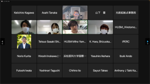
August 3rd, 2022, we had 14th journal club with Dr. Robert V. Warren who is a senior associate of Hamamatsu Photonics. For the sake of infection prevention of the COVID-19, it was held online.
In this time of journal club, 4 students from Shizuoka University and Hamamatsu University School of Medicine made a presentation about a research paper and discussed with the audience. The article of choice was one published in Journal of Otolaryngology Head Neck Surgery, with titled “Otitis Media Middle Ear Effusion Identification and Characterization Using an Optical Coherence Tomography Otoscope”.
The article discusses the evaluation results of an OCT device that was developed by a company PhotoniCare to diagnose otitis media. OCT has been widely used in the field of ophthalmology because of an ability to capture cross-sectional images of retina. In this article, the authors applied OCT to diagnosis of otitis media. The device demonstrated more than 90% of accuracy, sensitivity, and specificity*1 in terms of detection of liquid inside of inner ears. In addition to these results, the outcome is independent on medical experiences of its users. However, when it comes to determine whether the detected liquid is serous or non-serous, it gave 70% of accuracy, 53% of sensitivity, and 80% of specificity, which requires further improvement. Not only the diagnostic performances, but the usability was also tested and discussed the future improvements.
Ear consists of external ear, middle ear, and inner ear. Otitis media is a common disease among children compared to adults. This is because of structural difference in ear of children and adults. Eustachian tube of children is thicker and shorter than that of adults, which makes it vulnerable against infection of bacteria or other pathogens. Otitis media is classified into two types depending on its pathogenic mechanisms. One is acute otitis media that is caused by bacterial infection. The symptoms of this type are change in the color of tympanic membrane, its intumescence, perforation, and pain. Another is otitis media with effusion. This is induced by hyperactivity of eustachian tube. The symptoms of otitis media with effusion are formation of bubbles in tympanic membrane, its regression, and hearing loss. It is reported that it leads difficulty in language acquisition for children. A therapeutic method for otitis media with effusion is insertion of tympanostomy tube into tympanic membrane. To diagnose otitis media, otoscope or tympanometry is used. However, this diagnosis requires experiences for the physicians, and it scares and pains children making the diagnosis process difficult. With these difficulties, more simple and less invasive way to diagnose has been required.
Authors proposed to utilize Optical Coherence Tomography (OCT) to diagnose otitis media and evaluated its applicability. OCT is a real time imaging technique and is widely used in the field of ophthalmology to capture a cross-sectional image of retina. What makes it possible for OCT to visualize structures in its depth direction, is the fact that it is based on Michelson interferometer. Interference pattern obtained by the interferometer changes as per the travel distance of light that eventually interfere together and form the pattern. From its pattern, we can understand the travel distance of the light, in other words, the location where the light reflected from. Figure 1 [1] shows a device that the authors developed and is called “OtoSight”. They combined their OCT and conventional otoscope which keeps its design over the last 100 years. They aim to support clinician’s decision and reduce the invasiveness of its diagnostic processes.
To test the applicability of OtoSight, clinical trial for 70 children who receive tympanostomy tubes, was conducted. 20 OCT images were selected from 65 images taken from the 70 patients. These 20 images consist of 7 images of otitis media without effusion and 6 images of serous otitis media, 6 images of non-serous otitis media. Using the 20 images together with their duplicated mirror 20 images, readers were asked to diagnose depending on the images. The readers were 6 otolaryngologists, 6 pediatricians, 6 physician extenders*2, and 6 non-medical experienced people. The non-medical experienced people were recruited as a control group to evaluate the dependency on medical experiences. These 24 readers were classified into two groups depending on whether they have OCT experiences or not.
As a result, more than 90% of accuracy, sensitivity and specificity were achieved to determine the presence of effusion. It was shown that there was no correlation between the diagnostic result and OCT experience. Sensitivity and specificity depended on medical expertise. On the other hand, when it comes to determine whether the otitis media of interest is serous or non-serous, 70% of accuracy, 50% of sensitivity and 80% of specificity was recorded. Dependence on OCT experience was not observed in this case, either. Dependency on medical expertise was confirmed in accuracy and sensitivity.
Apart from the diagnostic results, regarding the fact that authors only obtained 65 readable images from 70 patients, the average age of 45 patients from whom authors acquired the 45 images, was 5.01 and that of 25 other patients was 2.54. The rendering ratio of readable images out of all the OCT images captured increased as clinical trial was going. The ratio was 50% in the first 6 months, it elevated to 63.6% in the next 6 months, and ending up being 69.4% in the next 6 months.
Based on the results, 5 improvement ideas are proposed. First is hardware improvement that makes the shape similar to the conventional otoscope. In the current design, the imaging component and the projection component are separated as shown in Fig. 1. The authors proposed to integrate these together as shown in Figure 2 [2], so the physicians feel familiar to OtoSight. Second is implementation of machine learning to differentiate serous otitis media from non-serous otitis media. To implement machine learning, it is required to prepare dataset for training its differentiate ability. For the dataset, it is important to relate images to quantitative analysis such as viscometrical measurement of the effusion. Third is to introduce a function that evaluate the readability of acquired images with OtoSight. Physicians take images of ear canal mistakenly as if it was a tympanic membrane. This is because of the physicians are unfamiliar with to identify tympanic membrane in OCT images. The fact that depth visualized by current OtoSight is shallower than conventional otoscope, makes it difficult to take a picture of tympanic membrane. One of the possible solutions is to use contrast to evaluate the readability. Fourth is re-designing the device based on ergonomics. Fifth is improvement of the user interface. By applying these five improvements, authors are aiming to apply OCT technology for otitis media diagnosis.
In the Q&A session, discussions shown below were made:
Question: While you explain about OCT, you mentioned that the visualized depth reaches more than 10 mm. What kind of situation are you assuming? I think this is too much.
Answer: I assume a situation of low scattering and absorption and using longer wavelength light. Imaging retina should be similar to this situation. The thickness of eyeball is somewhat like this.
Comment: Pursuing spatial resolution in depth direction sacrifices lateral resolution. This trade-off relationship is an important issue to overcome.
Question: When it comes to image structures in a depth direction, ultrasound imaging is available. The principle of this modality is based on measuring time in which the sound traveled in samples and estimate the depth. How does OCT calculate the depth? In terms of principle of depth estimation, what is the difference between OCT and ultrasound imaging?
Question: In the results part, you said that it was difficult to obtain readable images from younger children. Why is that? Is it something to do with the anatomical characters among children?
Answer1: I think this should be something to do with anatomical characters.
Answer2: I’ve heard that children move a lot, and it makes difficult to take readable images.
In this journal club, six students who take medical engineering program of Graduate school of Integrated Science and Technology in Shizuoka university, participated and asked following questions:
Question: What is ANOVA analysis?
Answer: ANOVA is an extended version of two sample t test*3. In the clinical trial, we cannot apply two sample t test since we have more than two groups (otolaryngologists, pediatrician, physician extenders, etc.) to compare.
Question: In the article, some patients were eliminated from the trial due to “sensory problems”. What are these problems?
Answer: I cannot provide you a clear answer since no details are provided in the article. In my opinion, the diagnosis is painful, so some might have declined to have the test.
Question: I cannot see differences between Figure 2(B) and (C) in the article. What exactly is the difference?
Answer: In Figure 2(C), the OCT signal is stronger than obtained in Figure 2(B). This implies that there are strong scattering substrates something like a solid rather than liquid. Although the two figures are different, it is difficult to discern in case of lower readability. In this regard, I think they need improvements something like machine learning implementation.
Question: If a patient is on anti-biotics that absorb OCT probe light, is this OtoSight still usable?
Answer: I think it is. We need to check the absorption wavelength of that anti-biotics beforehand. Even in the cases of strong absorption, once we clean the ear, I think we can avoid this issue.
Question: Apart from the ophthalmology and otolaryngology, is there any other field in which OCT is applied?
Answer: It is applied in cardiology, too. There is an application to image plaque*4. Besides the medical fields, semiconductor industry uses this. R&D is going about OCT in academia, too.
Question: What kind of technologies are required to improve OCT further in the future?
Answer 1: It is important to improve its scanning rate*5. Conventional OCT imaging takes time to render a 2D image due to its usage of line scanning.
Answer 2: It costs money, but spectroscopic analysis should be a good candidate to introduce. Current OCT cannot obtain chemical information about the scanned substrates, but spectroscopic analysis can.
Comment: It should be. In the field of cardiology, people would like to understand the chemical composition of the plaque.
Answer 3: In my thoughts, removing speckle noise*6 is important. Spatial resolution is good enough. Spectroscopic analysis is important to introduce. High speed imaging should be achieved using Full field OCT*7 after the improvement of its sensor.
Answer 4: I think combining OCT with other technologies or making the device smaller are important. We can integrate optical components of OCT in a way of integrated photonics*8.
Question: I am interested in companies that produce OCT devices like PhotoniCare. Do you know any other?
Answer: I cannot give you an immediate answer. You can search in the Web site of FDA.
Lastly, I, a writer of this report, make my comment on this journal club. I think the authors need to improve the device, especially about the determination of serous otitis media or non-serous one. But the issues to overcome is clear to tackle on in the future work and should be improved. I think to reduce the invasiveness and to deal with children’s movement are important problems to work on. It is easy to have a nice result for publication by recruiting patients who are quite susceptible to pain with zero movement, but engineering research should deal with more realistic situation so people can use the developed technology. I felt the collaboration of the engineering and medical science is a good opportunity for engineers to face such a practical point of view and should be encouraged more.
A small glossary
1: Accuracy, Sensitivity, Specificity
In general, these terms are used to characterize the performance of devices or measurements. Each of the fields applied, the definitions are different, which is the case for in this clinical study.
In a web page of CLESCO Ltd. (https://www.cresco.co.jp/blog/entry/5987/ Accessed:2022/09/05), which is a commercial company that supports ophthalmology, the definitions are described as below. Note: These are defined for the sake of practical applications.
Accuracy: An index to correctly determine positive or negative of a disease. (true positive + true negative) / total cases.
Sensitivity: An index to determine positive in case of a patient have a disease. true positive / (true positive + false negative).
Specificity: An index to determine negative in case of a patient do not have a disease. true negative / (false positive + true negative).
2: Physician extender
A professionality in the USA. Someone who can execute medical treatment under the supervision of a medical doctor.
Reference:英和生命保険用語辞典
3: Two sample t test
A statistical method that evaluates significant difference between two averages obtained from two independent populations.
Reference:統計Web (https://bellcurve.jp/statistics/course/9427.html), (https://bellcurve.jp/statistics/glossary/809.html) Accessed:2022/09/05
4: Plaque
A protrusion which is formed inside of an artery. As it grows in its size, it suspends blood flow.
Reference:いしまる内科クリニック(https://www.ishimaru-naika.com/policy20.html) Accessed:2022/09/05
5: Scanning rate
Frequency of laser scanning on a sample surface. It is possible to take an image with shorter period of time using high frame rate.
Reference:画像検査.com (https://gazou-kensa.com/camera/136/) Accessed:2022/09/05
6: Speckle noise
Point like noises on an image that is originated from interference of reflected illuminated laser.
Reference:Optipedia (https://optipedia.info/laser/handbook/laser-handbook-7th-section/31-3/) Accessed:2022/09/05
7: Full field OCT
A variation of OCT techniques that uses plane illumination and photodetectors aligned two dimensionally.
Reference:視覚の科学 (https://www.jstage.jst.go.jp/article/jpnjvissci/29/2/29_29.58/_pdf/-char/ja) Accessed:2022/09/05
8: Integrated photonics
A trend of photonics research that develops applications using integrated optical components on a single tip, which is similar to a way of conventional integrated circuit.
Reference: AIM Photonics (https://www.aimphotonics.com/what-is-integrated-photonics) Accessed:2022/09/05
References:
[1] Otolaryngol Head Neck Surg. 2020 March; 162(3): 367–374. doi:10.1177/0194599819900762
[2] Medical expo, (https://www.medicalexpo.com/ja/prod/syncvision-technology-98009.html) Accessed:2022/09/05
innovative Photonics Evolution Research Center (iPERC)
3-5-1 Johoku, Chuo-ku, Hamamatsu, Shizuoka 432-8011 Japan
phone: +81-53-478-3271 / fax: +81-53-478-3256
![Figure 1 Appearance of OtoSight [1]](https://www.iperc.net/en/mgr/wp-content/uploads/2022/12/Figure1-266x300.png)
![Figure 2 Coupling the imaging component with the projection part [2].](https://www.iperc.net/en/mgr/wp-content/uploads/2022/12/Figure2-300x282.png)
