

Report on the 15th iPERC Seminar
- 2023/03/10
On December 7, 2022, Dr. Robert Virgil Warren of HAMAMATSU VENTURES USA held an online Journal Club.
In this Journal Club, students from Hamamatsu University School of Medicine and Shizuoka University selected an academic article, divided their roles, gave a presentation, and discussed it with Dr. Warren and the participants. The presenters were Chihiro Kawabata, 4th year medical student, Hamamatsu University School of Medicine; Mayuko Kojima, 4th year medical student, also from Hamamatsu University School of Medicine; Tomohiro Okuyama, 1st year doctoral student, Cooperative Major in Medical Photonics, Shizuoka University; and ARA IFAT, 3rd year doctoral student, Graduate School of Science and Technology, Shizuoka University.
The Journal Club focused on the paper entitled “Dual-modality fluorescence lifetime imaging-optical coherence tomography intravascular catheter system with freeform catheter optics” [1]. In this paper, the authors proposed a multimodal imaging system that utilizes optical coherence tomography (OCT) and fluorescence lifetime imaging (FLIm) to assess the spatial structure and biochemical properties of intravascular vessels simultaneously. The application of this imaging system is expected to provide a deeper understanding of the pathophysiology of intravascular plaques and enhance the diagnosis of cardiovascular diseases.
Summary of Presentation:
Chihiro Kawabata, a fourth-year medical student at Hamamatsu University School of Medicine, discussed the formation mechanism of atherosclerosis and the significance of imaging them. Imaging techniques for atherosclerosis, such as coronary angiography and intravascular imaging, were explained. She also noted that intravascular imaging methods like intravascular ultrasound (IVUS) and optical coherence tomography (OCT) have a limitation in clarifying the composition of plaque*1. She suggested that fluorescence lifetime imaging (FLIm) can be an effective technique to determine the composition. Finally, she discussed the benefits of a new system that combines OCT and FLIm (FLIm-OCT).
Tomohiro Okuyama, a first-year doctoral student at Cooperative Major in Medical Photonics, Shizuoka University, explained the configuration and principle of FLIm-OCT. He described how multispectral fluorescence lifetime imaging can visualize many fluorescent dyes, such as collagen and elastin. Based on the schematic of the optical setup, the arrangement of FLIm and OCT and the principle of operation were explained. He explained about the catheter, which is an important module for intravascular imaging. The mechanism and performance evaluation results of the catheter were explained. Finally, he discussed how human coronary artery specimens are prepared and evaluated for use as samples for intravascular imaging.
Mayuko Kojima, a fourth-year medical student at Hamamatsu University School of Medicine, explained two main results from the paper. First, she discussed the evaluation results of a fiber optic rotary collimator (FORC), showing the amount of beam shift when the FORC is rotated. The stability of the system was demonstrated based on the small variation in the coupling efficiency*2 of the light. Second, she presented the results of arterial imaging, showing the spatial correspondence between the images taken by FLIm-OCT and those stained by Movat’s pentachrome*3 and CD68*4.
ARA IFAT, a third-year doctoral student at the Graduate School of Creative Science and Technology, Shizuoka University, discussed the catheter mounting and design of the rotation mechanism, semiconductor detector in the motor drive unit, and imaging results of arteries. In designing the catheter, she compared the conventional method using a ball lens with the method using free-form reflective optics used in this paper. It was demonstrated that the use of free-form reflective optics can improve the collection efficiency of the FLIm signal. The evaluation results of the beam shift and coupling efficiency showed that the proposed rotation mechanism has high adaptability and stability. Imaging results of arteries showed that macrophage form cells (mFCs) lead to changes in the fluorescence lifetime and an increase in the OCT signal. Finally, she presented prospects that combining spectroscopy and OCT can improve the detection and quantification of inflammation and improve methods for assessing the extracellular matrix.
Paper Outline:
Intravascular imaging represents an effective technique in the investigation of atherosclerotic plaques and the diagnosis of cardiovascular diseases. The authors of the paper have designed and characterized a multimodal imaging system that combines optical coherence tomography (OCT) and multispectral fluorescence lifetime imaging (FLIm). This system is capable of simultaneously assessing both the structural and biochemical properties of coronary arteries.
As illustrated in Figure 1, the optical axes of OCT and FLIm are aligned in the imaging system, and each light is introduced into a catheter. The light emitted from the catheter is directed to the affected area, and the reflected light is subsequently returned to the catheter. The catheter is rotated by a rotary collimator in the motor drive unit (MDU), which enables the acquisition of three-dimensional information about the blood vessel.
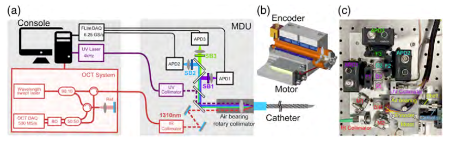
Fig. 1[1] (a) illustrates the schematic of the FLIm-OCT system, which comprises a catheter, motor drive unit (MDU), FLIm, and OCT modules. (b) shows a 3D model of the rotary collimator, with an encoder that reads the angular position of the rotary axis, enabling closed-loop control of the rotary axis and image reconstruction.
The authors of the paper proposed a FLIm-OCT catheter system, which includes several unique devices:
(1) A fiber optic rotary joint with air bearings
(2) A catheter system equipped with broadband free-form reflective optics (previously described in detail in a paper by the same authors [2])
(3) Semiconductor detectors for multispectral FLIm
Through system evaluation, the authors achieved excellent coupling efficiency and stability in the UV and IR regions (IR: 75.7% ± 0.4%, UV: 45.7% ± 0.35%), high optical performance for FLIm (FWHM for UV: 50 μm), and excellent beam quality for OCT (FWHM for IR: 17 μm) simultaneously. Additionally, high-quality FLIm-OCT images of human coronary artery specimens were obtained, as depicted in Figure 2. The obtained images were compared with optical images acquired by Movat’s pentachrome staining and CD68 staining. The comparison revealed that (a) areas with short fluorescence lifetime corresponded to healthy regions, while areas with long fluorescence lifetime corresponded to pathological intimal thickening (PIT*5) and early fibroatheroma (eFA*6), and (b) areas with long fluorescence lifetime corresponded to accumulated foam cells.
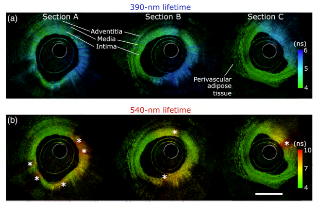
Fig. 2[1] Imaging results of a human coronary artery specimen. Brightness corresponds to backscatter intensity of OCT signal, and the hue represents the lifetime averaged by FLIm. (a) and (b) correspond to the 390 nm and 540 nm spectral bands, respectively. The depth information obtained through OCT facilitates the visualization of the whole vessel wall in healthy regions, including the perivascular adipose tissue.
The proposed FLIm-OCT system holds great promise for elucidating plaque pathophysiology and enhancing cardiovascular diagnosis by providing concurrent assessments of plaque structure and biochemical attributes.
Following each presentation, a Q&A session was conducted as follows:
Q1. Dr. Warren asked, “Are there any difficulties in constructing the FLIm-OCT system?” The presenter answered, “Optical construction of the catheter system poses a challenge. Unlike conventional OCT systems that employ a single wavelength, the proposed system utilizes a wide range of wavelengths. Hence, selecting an appropriate fiber for such a wide range is a difficult task.”
Q2. Dr. Warren asked, “Are the results of the FORC (fiber optic rotary collimator) evaluation good or bad?” The presenter answered, “Yes, it is the anticipated outcome. I know why the beam shift occurred.” Dr. Warren replied, “The shift is small, and the coupling efficiency is high. The coupling efficiency remains high and stable even when the catheter is rotated, which is a desirable outcome.”
Q3. An audience asked, “The coupling efficiency of FORC is stable, but what about unstable conditions?” Dr. Warren replied, ” What if there are vibrations in the operating room? The results presented here are from clinical trials and not actual use by physicians in the operating room. The coupling efficiency may fluctuate based on the physician’s movements and other unexpected situations. This aspect will be investigated in the future.”
Q4. An audience asked, “Does the laser light energy have any effect on the cells?” The presenter replied, “I think the effect is minimal because the light’s incident time is only 1 ns.”
Q5. Dr. Warren asked, “What happens during the interaction between light and tissue?” The presenter replied, “The fluorescence and scattering effects are significant. They appear as FLIm and OCT signals, respectively.”
Q6. An audience asked, “What is the penetration depth of FLIm light?” Dr. Warren answered, “The depth is 150 μm, which is shallow compared to the depth obtained by OCT.”
Q7. An audience asked, “Blood flows through actual blood vessels. How does this affect the results?” Dr. Warren indicated, “This issue was addressed in the frame rate discussion.” The presenter replied, “The sampling frequency is 6.25 GHz.” Dr. Warren asked, “What limits the frame rate? The presenter replied, “The rotation speed of the rotation mechanism affects it.” Dr. Warren replied, “The repetition rate also limits it. By using a light source with a higher repetition rate, a higher frame rate can be obtained. This will enable us to visualize blood flow and other complex motions.”
Q8. An audience asked, “The FLIm images are not as clear as the optical image. Do you have any ideas to obtain a clearer image in depth?” The presenter replied, “Pulsed laser irradiation with higher intensity would be good.” Another audience replied, “The images correspond to topography*7 and not tomography*8, so there is no depth resolution.”
Q9. An audience asked, “Do you have ideas to obtain depth resolution?” Dr. Warren answered, “It is very challenging because FLIm uses ultraviolet light, which has a short penetration depth, making it difficult to obtain depth information.” An audience indicated, “There are some reports using two-photon microscopy, but it is difficult to apply to catheters.” Dr. Warren suggested, “We had better consider near-infrared spectroscopy or other spectroscopic methods.”.
Q10. Dr. Warren asked, ” What is something you have learned with interest?” The presenters replied, “Understanding this paper was very difficult. I was not familiar with imaging technology, but I learned that it is wonderfully compatible with medical care. I would like to study it more in the future.”, “I am interested in the scan method of light.”, “I had no knowledge of fibers. I was surprised that we could use couplers to split and stack light.” Finally, Dr. Warren said, “The rotation mechanism, the coupling of two different systems, and the free-form reflective optics are very complex. I learned a lot.”
Reporter’s Comments:
The Journal Club focused on OCT-related research, building on the previous Journal club [3]. In the selected paper, OCT is utilized to obtain tomographic information on plaques that occur in blood vessels. While OCT has become a standard technique in ophthalmology, its application to intravascular imaging remains limited, and additional reports are necessary. In addition to the tomographic information obtained by OCT, the authors have also obtained tomographic composition information using FLIm. I was impressed with the implementation of the system as well as the usefulness of the acquired images. Simply by examining the optical setup shown in the paper, I sensed the difficulty and complexity of the implementation. The proposed FLIm-OCT system is not limited to intravascular imaging, but can be applied to fields such as ophthalmology, otolaryngology, and the semiconductor industry, and I consider it to be a subject worth keeping a close eye on in the future.
During the journal club, students and professors engaged in a lively discussion of the proposed method. As an engineering major, I was unfamiliar with the cases that appeared in the paper, but my questions were addressed during the student’s presentation and the discussion with Dr. Warren. This was an excellent opportunity to bridge the gap between engineering and medicine.
Finally, I would like to express my gratitude to Dr. Warren for leading the journal club, the students for preparing the presentation materials, and the individuals at Shizuoka University and Hamamatsu University School of Medicine.
References:
[1] Li C, Bec J, Zhou X, Marcu L., “Dual-modality fluorescence lifetime imaging-optical coherence tomography intravascular catheter system with freeform catheter optics,”J Biomed Opt., 27(7), 076005 (2022).
[2] J. Bec et al., “Broadband, freeform focusing micro-optics for a side-viewing imaging catheter,” Opt. Lett. 44, 4961-4964 (2019).
[3] “第14回iPERCセミナーを開催しました”, 光創起イノベーション研究拠点, 2022-10-11 https://www.iperc.net/news/20220803journalclubreport/
Grosarry:
- Plaque: An accumulation of foam cells, resulting from the oxidation of LDL, which accumulates in the intima of blood vessels and contributes to the development of atherosclerosis.
- Coupling efficiency: The ratio of light intensity taken into the fiber to the light intensity incident on the fiber. Because light travels by total reflection within the fiber, the fiber material must be optimized for the light wavelength. In this paper, light with a wavelength range from UV to IR is used, necessitating the selection of a fiber with a wide bandwidth.
- Movat’s pentachrome: A stain used in the study of vascular diseases that can stain collagen, elastin, muscle, and other tissue components.
- CD68: An immunohistochemistry used in selected paper to specifically detect macrophages.
- PIT (pathological intimal thickening): A lesion consisting of macrophages and extracellular fat, representing an early stage of atheroma formation.
- eFA (early fibroatheroma): Referring to a plaque with a fibrous structure.
- Topography: ‘topo’ meaning ‘place’. In the fields of electron microscopy and optical measurement, it refers to the three-dimensional shape of a sample surface.
- Tomography: ‘tomo’ meaning ‘cut, incision’. Computed tomography (CT) is a well-known method of obtaining tomographic information by computer processing.
innovative Photonics Evolution Research Center (iPERC)
3-5-1 Johoku, Chuo-ku, Hamamatsu, Shizuoka 432-8011 Japan
phone: +81-53-478-3271 / fax: +81-53-478-3256