
第10回iPERCセミナーを開催しました
- 2020/06/08
2020/05/18 The 10th iPERC Seminar (The 6th Journal Club)
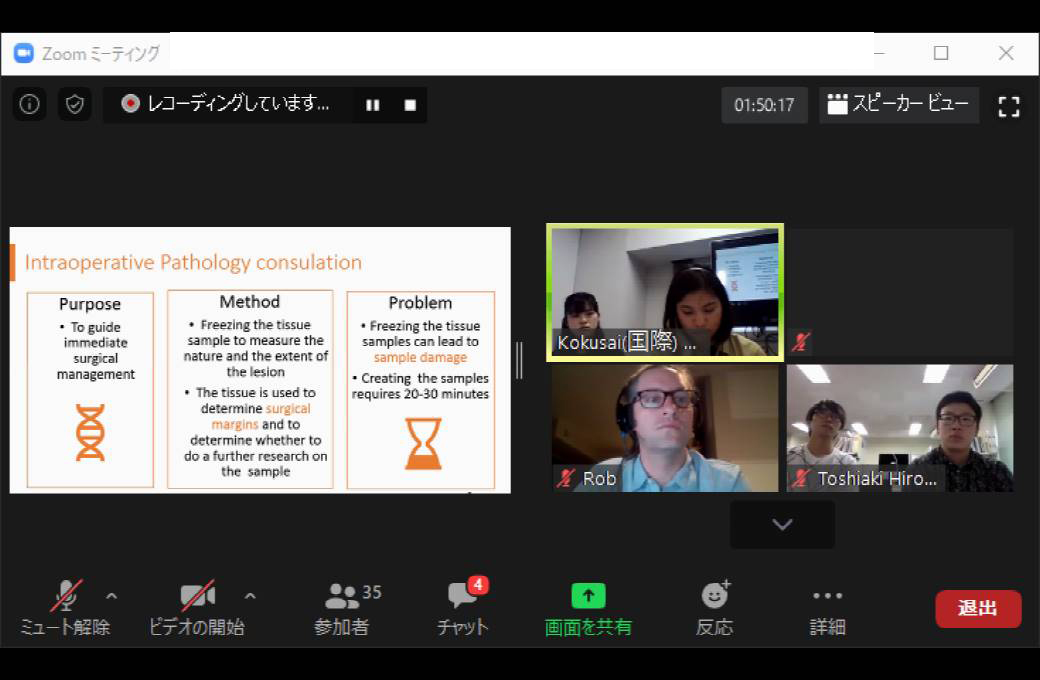
We held the 6th Journal Club with Dr. Robert Virgil Warren, who is a project scientist at the Beckman Laser Institute, on May 18. To prevent the spread of COVID-19, the journal club was a video conference. 35 people participated.
In this journal club, 2 students at Shizuoka University and 2 students at Hamamatsu University School of Medicine broke down an article into parts and made presentations. ‘Light-sheet microscopy for slide-free non-destructive pathology of large clinical specimens’ was selected as the article.
In the article, current methods for pathological specimen examination were discussed and the authors proposed new methods involving light-sheet microscopy.
In the US, treatment decisions for the 1.7 million annual patients who receive a new cancer diagnosis are largely based on histopathological specimen examinations. The gold standard of slide-based microscopic pathology suffers from high inter-observer variability and limited prognostic*1 value due to sampling limitations and the inability to visualize tissue structures and molecular targets in their native 3D context.
While 3D digital pathology using serial sections on glass slides is possible, it is not a viable strategy for routine clinical use due to the labor and time involved in preparing and imaging hundreds of glass slides, and aligning the digital images. Non-destructive fluorescence microscopy of human tissues has been attempted using a variety of approaches. However, each of them has a weak point such as imaging depth, complexity, phototoxicity or imaging speed.
The article proposed open-top light-sheet microscopy, which provides low phototoxicity, large imaging area, and adjustable imaging speed to suit specimen depth.
Light-sheet microscopy is a kind of fluorescence microscopy. This technique illuminates a light sheet into a specimen and detects the fluorescence from a focal plane along a different axis than illumination to minimize fluorescence from out-of-focus features. 3D digital pathology can be acquired by imaging while moving the specimen and focal plane. As compared with epifluorescence microscopy, light-sheet microscopy can acquire images in focus with low phototoxicity by only illuminating the focal plane. However, this technique has a limitation of specimen size because optical system was located close to specimen.
In the proposed method, an open-top architecture can image larger specimens because the optical system is located under the microscope stage and there is no limitation with moving the specimen. To enable aberration*2-free imaging, this method uses a hemispherical SIL*3.
A variety of clinical pathology applications were explored. These experiments demonstrate that the proposed method is optimal and versatile for both rapid microscopy of irregular tissue surfaces and comprehensive volumetric microscopy.
Questions and answers are as follows.
Q1: What is the magnification of the proposed method?
A1: The spatial resolution is 1.4 µm. It can image very small structures.
Q2: Technically, what is the factor limiting the imaging speed?
A2: Imaging speed is determined by the frame rate of camera. Frame rate is limited by the specimen’s height because large image heights need a large crop size.
Q3: Two objective lenses are placed under the microscope stage. Probably, objective lenses interfere physically if they are big. Are there any special designs for objective lenses or any limitations about lens’ NA and working distance?
A3: The SIL effectively increases the illumination and detection NA, which are both quite small without the SIL (NA = 0.03 for the light sheet and NA = 0.28 for the detection objective lens). As a result, for both ends of the optical setup the working distance is large and there are not physical limitations.
Q4: Using a conventional approach for pathology, you can look at specimens retrospectively because they were homogenized and stored. How does the proposed method keep specimens?
A4: The students were uncertain of how to keep specimens which were prepared for light-sheet microscopy, but during discussion with Dr. Warren it was pointed out that it’s possible to virtually re-analyze samples because volumetric data is acquired and stored.
Dr. Warren’s question: The first presentation of light-sheet microscopy was all the way back more than 100 years ago. Why is light-sheet microscopy popular right now? What types of things have changed over the last 10 or 15 years that suddenly light-sheet microscopy is getting very popular?
Student answer: In recent years, living specimens have been imaged in neuroscience and brain science. I think light-sheet microscopy’s low phototoxicity is a big advantage compared to conventional microscopy.
Dr. Warren’s supplementary explanation: It definitely has become a popular tool for research, but there are also some big technology developments. There’s been advancements in CMOS cameras over the last 10 years that have been very important and they’ve kind of been an enabling factor. It suddenly changed the game.
In addition to the above questions and answers, there were discussions about experimental conditions and diagnosis by artificial intelligence.
In the article, it is discussed that there is great opportunity with light-sheet microscopy and information technology such as computer-aided diagnosis and automated image interpretation.
It is expected that there can be improvement of imaging speed and image quality by improving the camera’s performance and data processing methodologies such as scatter correction.
Article
GLASER, Adam K., et al. Light-sheet microscopy for slide-free non-destructive pathology of large clinical specimens. Nature biomedical engineering, 2017, 1.7: 0084.
https://www.ncbi.nlm.nih.gov/pmc/articles/PMC5940348/
References
*1 prognosis: The forecast of the probable outcome or course of a disease; the patient’s chance of ‘recovery.
(MedicineNet. Definition of Prognosis, Retrieved June 13, 2020,
from https://www.medicinenet.com/script/main/art.asp?articlekey=5061)
*2 aberration: A property of optical systems that causes light to be spread out over some region of space rather than focused to point.
(Kirkpatrick, Larry; Wheeler, Gerald (1992). Physics: A World View (2nd ed.). Philadelphia: Harcourt Brace College Publishers. p. 410)
*3 solid immersion lens (SIL) : It extends the diffraction limit by filling the object space with a high refractive index material.
(WU, Qian; GHISLAIN, Luke P.; ELINGS, V. B. Imaging with solid immersion lenses, spatial resolution, and applications. Proceedings of the IEEE, 2000, 88.9: 1491-1498.)
---------------------------------
5月18日、Beckman Laser InstituteのProject ScientistであるRobert Virgil Warren先生をお迎えして、光創起イノベーション研究拠点のJournal Clubを開催しました。新型コロナウイルス感染症の感染防止のため、今回はWeb会議システムを用いたオンラインセミナー形式で開催しました。
本セミナーは、浜松医科大学と静岡大学の学生が1つの英語論文を役割分担して発表するJournal Clubです。今回は、「Light-sheet microscopy for slide-free non- destructive pathology of large clinical specimens」を題材にして、病理組織診断のために大きな検体をスライド不要で観察可能にする光シート顕微鏡法について議論しました。
論文では、顕微鏡による病理学的標本検査の現状と提案手法について次のように論じられています。
アメリカで新たに癌診断を受ける年間170万人の患者の治療法決定は、病理学的標本検査に基づいています。しかし、その至適基準: gold standardである顕微鏡による検査は、目的の組織をそのまま3次元で可視化することが困難なため、観察者により評価の変動が大きく、予後*1の推定精度に限界があります。
従来の手法では、人体から採取した検体を複数の薄い切片に加工してスライドガラスに封入し、撮像することで3次元情報を取得できます。しかし、作業工程が多いことにより時間と労力を要するため、臨床で他の業務に加えることは困難です。加えて、作業工程による検体の劣化や、撮像領域が小さいことから、評価のばらつきが大きいことが問題になっています。非破壊の顕微鏡法にさまざまなアプローチがされてきましたが、検体の損傷や、厚み方向の撮像可能領域、撮像速度等にそれぞれ欠点があります。
この論文の提案手法であるオープントップ型光シート顕微鏡法は、検体の損傷を抑えられる、検体サイズ(縦横幅)に制約がない、厚みに合わせて撮像速度を調整可能である、という特徴があります。
光シート顕微鏡法は、薄いシート状の励起光を検体側方から照射し、励起光と垂直方向に配置したカメラで焦点面の蛍光を検出する非破壊の顕微鏡法です。検体を移動して焦点面を移動しながら撮像することで、3次元情報を取得します。焦点面以外に励起光を照射しないため、通常の落射蛍光顕微鏡法と比べてピンぼけしづらく検体の損傷が抑えられます*2。ただし、光学系が被写体付近に配置されていることから、撮像可能な検体サイズに制約がありました。
提案手法では、光学系をステージ下面に配置し上面を開放することで、検体が自由に移動できるようになるため、大きな検体が撮像可能になりました。また、励起光と収集光がステージのガラスを透過する際に発生する収差*4を最小限に抑えるため、ステージ下面に薄い油層とソリッドイマージョンレンズ*3が配置されました。
臨床病理学的利用法が検証され、不規則な表面の検体を含む臨床検体の3次元イメージングと、表面イメージングの両方を迅速に行うことができることから、さまざまな用途で利用可能であることが示されました。
各発表者の発表が終わると、聴講者から発表者に対して以下のような質疑応答が行われました。
Q1: 提案された顕微鏡法ではどの程度まで拡大可能か。
A1: 1.4 µmの分解能がある。とても小さな構造をイメージングできる。
Q2:被写体の厚みに対して撮像速度が制限される技術的課題は何か。
A2: 撮像速度はカメラのフレームレートによって決まる。厚みが大きい被写体に対応してカメラのクロップ*5サイズを大きくするには、フレームレートを小さくする必要がある。したがって、厚みが大きい場合、フレームレートを小さくする必要があるため、撮像速度が小さくなる。
Q3: 光学系について作動距離: working distanceによっては対物レンズ同士が物理的に干渉することが考えられるが、何か特別な設計がされているのか、もしくはレンズの開口数に制約があるのか。
A3: 集光に用いるレンズは開口数が大きくSIL*3の近くに配置されているが、励起光の入射に用いるレンズは開口数が小さく作動距離が大きいため干渉しない。
Q4: 検体について、従来の手法では均質化してスライドガラスに封入することで、後から観察しなおせるが、提案手法で用いられたプロトコルの場合どのように検体を保存しているのか。
A4: 検体を染色する前にホルマリンを用いて処理している。どのように保存しているかはわからないが、データが保存されているためそれを見返すことができる。
Warren先生の補足: 3次元イメージングには利点があり、最初に検体全体をイメージングできていれば、再び顕微鏡で観察しなくとも、そのデータを参照して異なるスライドやセクションから観察することも可能である。
Warren先生の「光シート顕微鏡法は1世紀以上前からある技術なのに、なぜここ10数年で人気になったのか」という質問に対し、発表者は「脳科学等で生きた被写体が撮像されるようになり、光シート顕微鏡法は光毒性が小さいことが理由だと考えられる」と回答されました。Warren先生はその意見に同意し、「ここ10年のCMOSカメラの進歩はとても重要で、CMOSカメラの登場が、光シート顕微鏡法が有用であるという流れを作った」と補足されました。
上記の質問以外に、具体的な撮像条件に関する質問や人工知能による診断の話題などさまざまな議論が行われました。
論文では、複数のカメラを用いることでイメージング速度を向上可能であることや、コンピュータ支援診断システムおよび自動画像解釈の取り組み等の展望について論じられていました。今後、カメラの性能向上や散乱線補正等のデータ処理によって、より高速で高画質な3次元顕微鏡イメージングが可能になることを期待しています。
■題材論文
GLASER, Adam K., et al. Light-sheet microscopy for slide-free non-destructive pathology of large clinical specimens. Nature biomedical engineering, 2017, 1.7: 0084.
https://www.ncbi.nlm.nih.gov/pmc/articles/PMC5940348/(2020年5月20日)
■参考文献
*1 予後: 手術や病気、創傷の回復の見込みを意味する医学用語
(出典: 予後 – 薬学用語解説.日本薬学会.
https://www.pharm.or.jp/dictionary/wiki.cgi?予後(2020年5月28日))
*2 野中茂紀, 「光シート顕微鏡:生体観察のための新しい顕微鏡法 Light-Sheet Microscopy: A New Principle for Live Imaging」, 『顕微鏡』第47巻 3号, 2012, pp.163-166,
http://microscopy.or.jp/jsm/wp-content/uploads/publication/kenbikyo/47_3/pdf/47-3-163.pdf(2020年5月23日)
*3 ソリッドイマージョンレンズ(Solid immersion lens : SIL):半球型または超半球型をしており、空間分解能を空気中の回折限界以上にすることができる。底面の中央付近の像を球面収差なく結合可能であり、近接場顕微鏡に用いられる。
(出典: 秋山英文ら.「ソリッドイマージョンレンズ(SIL)を 用いた近接場蛍光顕微計測法.」, 『応用物理』第71巻6号, 2002, pp.716-717
https://www.jstage.jst.go.jp/article/oubutsu1932/71/6/71_6_716/_pdf(2020年5月28日))
*4 収差: 理想結合からのずれ。像がぼけ、歪む原因となる。
(出典: 加藤欣也, 「レンズ光学の基礎3: 光学系の収差」, 『視界の科学』第36巻 3号, 2015, pp.40-44)
*5 クロップ(crop):イメージセンサのうち中央部分のある範囲のみを使って撮影する機能。データ処理量が減るため、連写性能が向上する。
(出典: クロップとは.【ライカイズム】写真用語辞典.
https://leicaism.jp/glossary/crop.html(2020年5月28日))


- 2024/01/25
- 2023/11/17 HAMAMATSU異分野交流会を開催しました
- 2023/10/17
- 第16回iPERCセミナーを開催しました
光創起イノベーション研究拠点棟 〈光創起研究棟〉
〒432-8011 静岡県浜松市中央区城北3丁目5-1 国立大学法人静岡大学浜松キャンパス内
TEL:053-478-3271 / FAX:053-478-3256