

Report on the 11th iPERC Seminar
- 2021/03/26
On January 18, 2021, the Journal Club by Dr. Robert Virgil Warren, Project Scientist at Beckman Laser Institute & Medical Clinic, the University of California, Irvine, was held online.
In this Journal Club, students from Hamamatsu University school of Medicine and Shizuoka University had presentations about one academic paper which they selected. After that they discussed with Dr. Warren and audience. The presenters were Mr. Tatsuki Furuhashi, a 1st year graduate student in Graduate School of Integrated Science and Technology, Department of Engineering, Shizuoka University, Mr. Hoang Son Nam, a 1st year graduate student in Graduate School of Integrated Science and Technology, Department of Engineering, Shizuoka University, Mr. Tatsuki Masuda, a 5th year undergraduate student in the Faculty of Medicine, Hamamatsu University School of Medicine, and Mr. Yasuhiro Ikehara, a 3rd year undergraduate student in the Faculty of Medicine, Hamamatsu University School of Medicine.
In this journal club, an original paper entitled “Smartphone snapshot mapping of skin chromophores under triple-wavelength laser illumination”[1] by Janis Spigulis et al., University of Latvia was used as a material. We discussed the research that enables mapping of chromophores (melanin, hemoglobin, etc.) in skin tissue using snapshots from a smartphone camera.
Presentation outline
Mr. Ikehara, Hamamatsu University School of Medicine
Mr. Ikehara covered the basis in skin tissues such as dermis and epidermis, and skin lesions (nevus, pigmented seborrheic keratosis, hemangioma) that were dealt in the paper. He also covered the features and problems of currently used detection devices and the MSI (multispectral imaging) which is one of modalities using such devices and was tried to improve in the paper.
Mr. Hoang, Shizuoka University
Mr. Hoang covered the acquiring method of images of chromophores distribution in skin tissue and the selection of wavelengths used in the measurements. He also explained the light propagation in skin tissue and the Monte Carlo method used to estimate the propagation distance.
Mr. Furuhashi, Shizuoka University
Mr. Furuhashi covered the configuration of the illumination unit, which consists of a laser module, diffuser and battery, and explained the illumination method. He explained the experimental results of skin chromophores distribution acquired by the simultaneous uniform illumination of 3 wavelength lights, and raised problems about the speckle noise generated from the system and the effects of other chromophores that were not taken into account.
Mr. Masuda, Hamamatsu University School of Medicine
Mr. Masuda discussed the acquired images from a viewpoint of clinician. He summarized the information on chromophores (oxyhemoglobin, deoxyhemoglobin, and melanin) in the acquired images from nevi, pigmented seborrheic keratosis, and hemangioma lesions. In addition, he introduced the necessity of clinical application (to establish it as a gold standard) and improvement of the system so that anyone can handle it easily.
Abstract of the paper
Mapping images of the chromophores distribution (melanin, hemoglobin, etc.) in skin tissue can provide very useful information for the evaluation of skin lesions. For example, it allows qualitative and quantitative monitoring of skin conditions after burns, surgery, etc., thus enabling more accurate diagnosis.
There are various methods to acquire chromophores distribution, such as hyperspectral imaging (HSI) [2], spatial frequency domain imaging (SFDI) [3] and multispectral imaging (MSI) [4]. However, commercially available instruments for these methods have disadvantages such as long image acquisition time, high machine price, and the need for wired connections. Due to such difficulties, these instruments are difficult to use for the tailor-made medical care in various environments and situations including outdoor use and personal use.
Since simple and rapid measurements are desired in clinical practice, the authors tried to improve the MSI system. In that system, the authors simultaneously irradiated laser light at three wavelengths (448-532-659 nm) to the area of skin chromophores (melanin, oxyhemoglobin, and deoxyhemoglobin). The reflected light was captured as RGB image data by a smartphone camera. Figure 1 [1] shows an overview diagram of the system. As shown in this figure, it is a simple system configuration in which a smartphone is combined with a light source and an imaging system via an adapter.
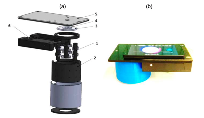
| Fig. 1[1] (a) The model diagram of the triple-wavelength illumination unit, (b) a smartphone attached with a prototype of this measurement unit. |
The features of this system are that it only requires a single snapshot and that it uses the camera installed in a smartphone as an imaging device which anyone can afford now. Advantages to use the smartphone include that the immediate processing of the acquired data can be done and that sending data to other devices to share is easy.
The first step in the measurement is to acquire three spectral images related to the incident wavelength. These images are processed according to Lambert-Beer’s law [5] to obtain the spatial increase or decrease of each of the chromophores. In addition, the mean free path of photons in the skin is estimated by Monte Carlo simulation.
In this paper, three types of skin lesions (nevi, pigmented seborrheic keratosis, and hemangiomas) were observed, and the spatial distribution of the increase or decrease of melanin, oxyhemoglobin, and deoxyhemoglobin was obtained. As an example, the measurement results of hemangioma are shown in Figure 2[1]. Figure (a) shows an RGB image. Figure (b-d) show oxyhemoglobin, deoxyhemoglobin, and melanin, respectively. These distributions are in good spatial agreement with the RGB image. In these images, the concentrations of oxyhemoglobin were increased, and that of deoxyhemoglobin were decreased, and that of melanin were unchanged. The result indicated that the concentration information of each chromophores was properly separated and extracted.
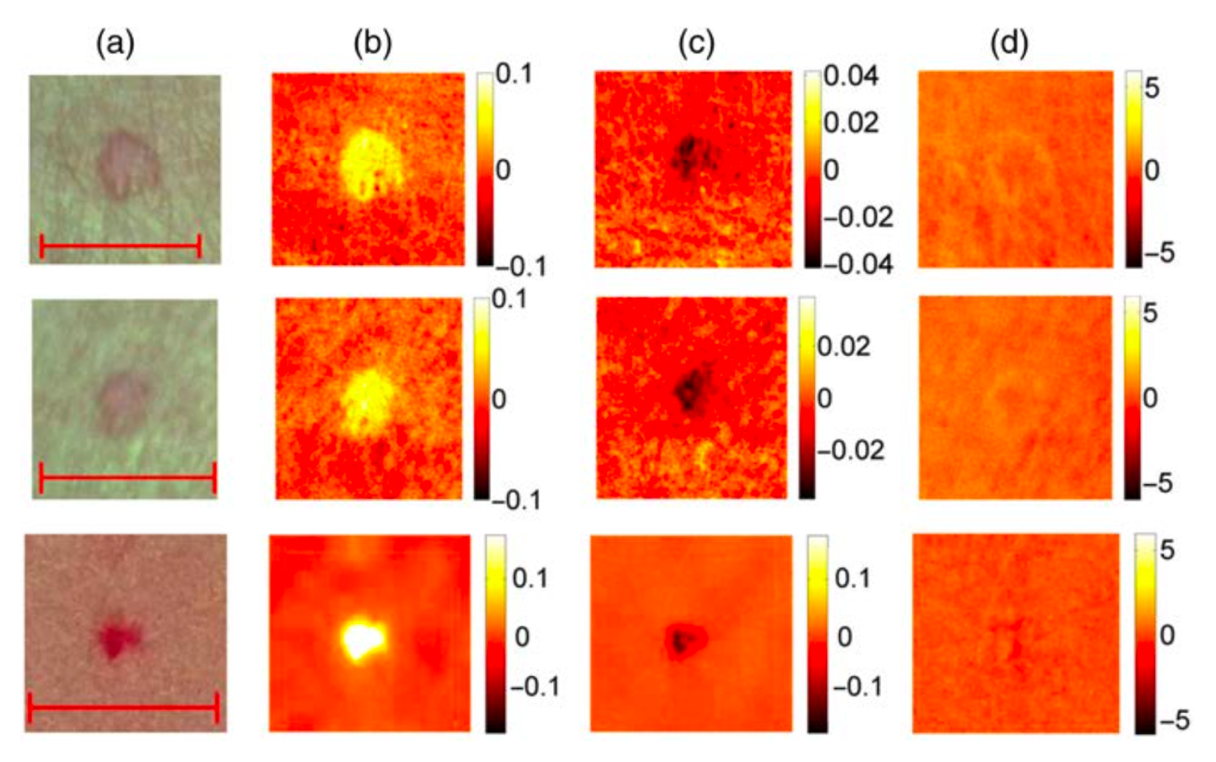
| Fig. 2[1] Imaging results of concentration changes of chromophores in 3 cases (top, middle, and bottom) of hemangioma (a) RGB image(scale bar: 5mm) (b)oxyhemoglobin (c) deoxyhemoglobin (d) melanin (units of color scale: mM) |
There are three issues that need to be addressed in the future: (1) reduction of speckle noise caused by a laser, (2) improvement of algorithms and hardware for image processing, and (3) increasing the number of chromophores that can be imaged [4].
After each presenter’s presentation, Dr. Warren and the audience asked the presenters the following questions.
Q1. A participant asked, “In this paper, three types of chromophores are considered. Are there any way to improve it so that we can see more than three types of chromophores in the future?”
Dr. Warren responded, “It would be good to refer the author’s previous paper and use more than three wavelengths.” Another opinion was given that if we add another light wavelength, we need to add another equation to the three equations described by the Lambert-Beer’s law.
Q2. Dr. Warren asked, “The authors did not distinguish nevi and pigmented seborrheic keratosis in the measurement. How will we be able to distinguish them?”
The presenter replied, “At this stage, there is little measurement data for nevi and pigmented seborrheic keratosis, so we need to conduct further measurements to clarify the difference.” Dr. Warren agreed with the opinion, and mentioned that AI could be applied to the data processing. Finally, he noted that nevi and pigmented seborrheic keratosis occur in different layers of skin tissue. He summarized that it would be possible to distinguish between nevi and pigmented seborrheic keratosis by using MSI and SFDI, which provide depth information.
Q3. A participant asked, “Is it possible to use IR or UV laser?”
The presenter answered, “IR imaging does not provide distribution information due to a low absorption of hemoglobin in the skin, and UV imaging does not provide depth information due to its shallow penetration depth into the skin. Therefore, the use of IR and UV laser in skin tissue observation is not appropriate.” Another participant stated, “In terms of detectors, the sensors in smartphones are sensitive only to visible light, which limits the detection range. Therefore, in order to expand the wavelength to UV and IR, we need to add new UV and IR cameras like those used in machine vision.”
Q4. A participant asked, “What are the advantages of using a smartphone as a measurement device?”
Another participant replied, “The advantage is that a smartphone makes telemedicine possible. In Japan, shortage of doctors is a serious problem in some areas, and telemedicine is one of the ways to solve this problem. By installing measurement devices in remote areas and connecting them to a doctor, patients will be able to receive a diagnosis even if the doctor is not there. For example, a patient can take measurements with a smartphone while at home. After the measurements, the data can be analyzed remotely, thus the participant can receive a diagnosis based on the results without going to a hospital.”
Q5. Dr. Warren asked, “Are there any weaknesses or points that need to be improved in the proposed method in this paper?”
A presenter replied, “The first weakness is that Monte Carlo simulation is required to calculate the photon path in the skin tissue. The accuracy of the Monte Carlo simulation is not always good. We proposed the time of flight method as a solution to this problem. The second weakness is that the authors analyzed only typical, more common diseases in this paper, which have a large number of cases. It is necessary to apply this imaging technology to more diverse cases with various diseases in clinical practice.”
Q6. Dr. Warren asked, “Is it possible to use LED and filters instead of laser?”
A presenter replied, “The authors introduced a research using LED in the introduction of this paper, so I think it is possible. ”Dr. Warren replied, “It is quite possible to use LED, and speckle noise can be removed by using incoherent light.”
Many other questions were raised, such as the medical usefulness of the measurement for nevi and pigmented seborrheic keratosis, and whether it is possible to estimate the depth of melanocytes. The lively discussion lasted for one hour.
Discussion with Dr. Warren after the presentation
Reporter’s comments
Smartphones are “computers” used by people all over the world now, for entertainment and business. They can also be used as medical devices as described in this paper. By using a smartphone as an imaging detector, the acquired data can be instantly shared without the need for expensive equipment. The accumulation of medical case data will facilitate applications in machine learning and deep learning thus enabling an accurate diagnosis of cases with complex pathologies and shapes of lesions. The improved data transmission speed will allow telemedicine without delays, which could help mitigate the effects of future doctor shortages.
Through the journal club, Dr. Warren, students and audience actively discussed the proposed method in the paper. Because I am majoring in engineering, I was not familiar with the medical cases presented in the paper. However, my questions were solved through the presentations and discussions with Dr. Warren. I think it was a very good opportunity to fill the gap between engineering and medicine.
Finally, I would like to thank Dr. Warren for teaching in the journal club, the students for preparing their presentations, and the people at Shizuoka University and Hamamatsu University School of Medicine.
References
[1] Janis Spigulis et al., Journal of Biomedical Optics, 22, 9, 091508 (2017). (J Biomed Opt. 2017 Sep 1;22(9):91508.doi: 10.1117/1.JBO.22.9.091508.)
[2] イメージ分光の原理/ハイパースペクトルイメージングとは Headwall Photonics | 特殊カメラ | 株式会社アルゴ(Last viewed date:February 3, 2021)https://www.argocorp.com/cam/special/HeadWall/how_it_works.html
[3]Amr Yafi et al., Lassers in Surgery and Medicine, 49, 827-834 (2017).
[4]福田弘之,堀江卓二,3-3 マルチスペクトルイメージングの産業応用,情報メディア学会誌,68(4),pp. 295-298 (2014)
https://www.jstage.jst.go.jp/article/itej/68/4/68_295/_pdf
(Last viewed date:February 3, 2021)
[5] 生体情報の取得原理 | スペクトラテック(Last viewed date:February 3, 2021)https://www.spectratech.co.jp/technology/bioloInfo.html
innovative Photonics Evolution Research Center (iPERC)
3-5-1 Johoku, Chuo-ku, Hamamatsu, Shizuoka 432-8011 Japan
phone: +81-53-478-3271 / fax: +81-53-478-3256